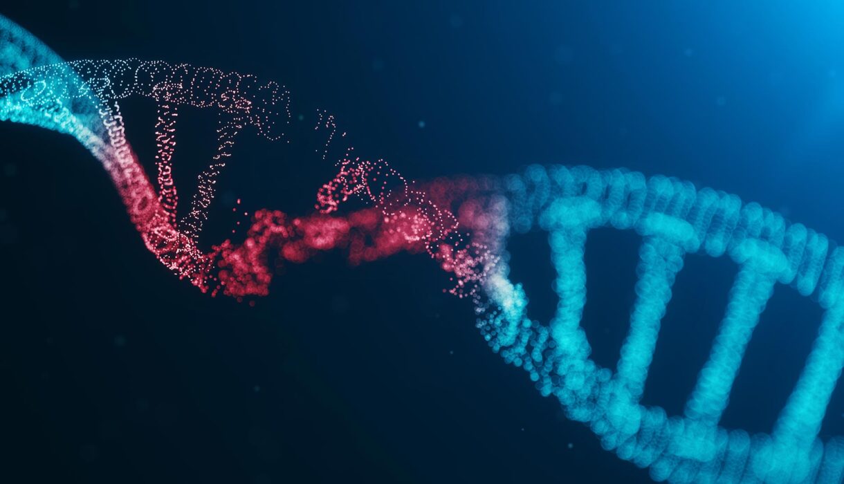
Imagine seeing the cell respond to damage as it happens: seeing breaks in its DNA, the arrival of repair proteins, and the very moment the damage disappears. Scientists at Utrecht University just made that possible. On November 23, 2025, the team reported a new fluorescent sensor: designed to track DNA damage and its repair, live and in real time, inside living cells and even whole organisms.
What Is This Sensor and How Does It Work
- The sensor is built using a small domain from a natural protein that cells already use.
- A fluorescent tag is attached to this domain. When DNA gets damaged, a specific marker appears on the damaged site, and the sensor binds, briefly and gently, lighting up that spot.
- Because the binding is reversible and non-disruptive, the sensor does not block the cell’s own repair machinery.
- In short: damage appears, the sensor lights up, repair proteins come in, and once the site is fixed, the sensor leaves. It gives a continuous movie of the repair process.
Why This Is a Big Deal
Before this, scientists studied DNA repair by taking “snapshots.” They would fix cells at different times, kill them, stain them, and piece together what happened. But this method misses a lot, it can’t show what happens in between those snapshots.
With the new sensor:
- Researchers can see the full repair sequence as it happens in living cells.
- They can track how fast repair proteins arrive, how long they stay, and when the DNA is fully restored.
- Because it’s gentle and real-time, it offers a more realistic picture of how cells behave naturally.
Beyond Cell Cultures: In Living Organisms
The team didn’t just test the sensor in lab-grown cells. They also tested it in C. elegans worms, a classic model organism for biology.
- In these worms, the sensor detected programmed DNA breaks, which are a normal part of development.
- This shows the tool isn’t just for petri dishes, it works in real, living animals.
Potential Applications
This sensor could revolutionize many fields:
- Cancer Research: Many cancer treatments work by damaging DNA. With this sensor, researchers can closely track how effective a drug is and how cells recover. It could make early drug testing more accurate, faster, and cheaper.
- Ageing Biology: DNA damage accumulation is linked to aging. Now, scientists can actually watch when and where damage happens and how well it gets repaired.
- Toxicology & Drug Safety: The sensor can help measure how much damage environmental toxins or new compounds cause, in real time.
- Basic Research: Because the sensor is modular (it can be connected to other molecular parts), scientists can map where damage happens in the genome, figure out which proteins gather at damage sites, or even move damaged DNA around in the nucleus to test hypotheses.
Why This Sensor Is Better Than Old Methods
- Non-invasive: The traditional tools, such as antibodies, bind too strongly to DNA and can disrupt repair. But this sensor binds gently and leaves, giving a more faithful signal.
- Continuous Imaging: Instead of numerous single experiments, one experiment now provides a complete timeline with more data.
- High resolution: Scientists can watch exactly when the repair proteins arrive, how many, and when they leave-all with very fine detail.
- Open access: The researchers made the tool freely available to other labs, meaning it could spread fast across the scientific community.
What Comes Next
- Mapping Damage: Scientists can now use the sensor to map where in the genome DNA damage happens most often.
- Understanding Repair Choice: By tracking which repair proteins come in, researchers can learn more about how cells choose different repair pathways (some are fast but error-prone, others are slow but accurate).
- Disease Study: The tool could help study diseases where repair goes wrong, like cancer or genetic disorders.
- Drug Testing: Pharma companies may use the sensor to test new therapies or to measure unintentional DNA damage caused by drugs.
Watch to know more
Conclusion
The new fluorescent sensor from Utrecht University allows for real-time observation in living cells of DNA damage and repair without interruption of the process. That is a huge step forward because old snapshot-based techniques are replaced by a true “live movie” of repair. The implications are immense: better testing of cancer drugs, deeper understanding of aging, and powerful basic research tools. In fact, as this technology spreads, we may soon understand our cells’ response to damage far better than ever before, and use that insight to design better treatments and interventions.